
Scoliosis Braces and Bracing
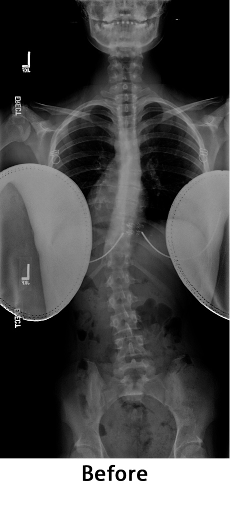
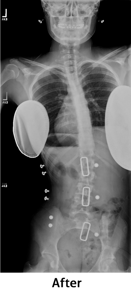
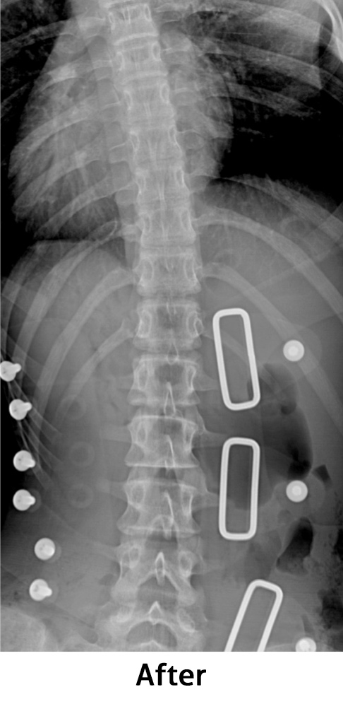
This patient received an Owens Carolina Nocturnal Brace. She is 14 years old. She presented with a pre-brace left curve of 28 degrees.
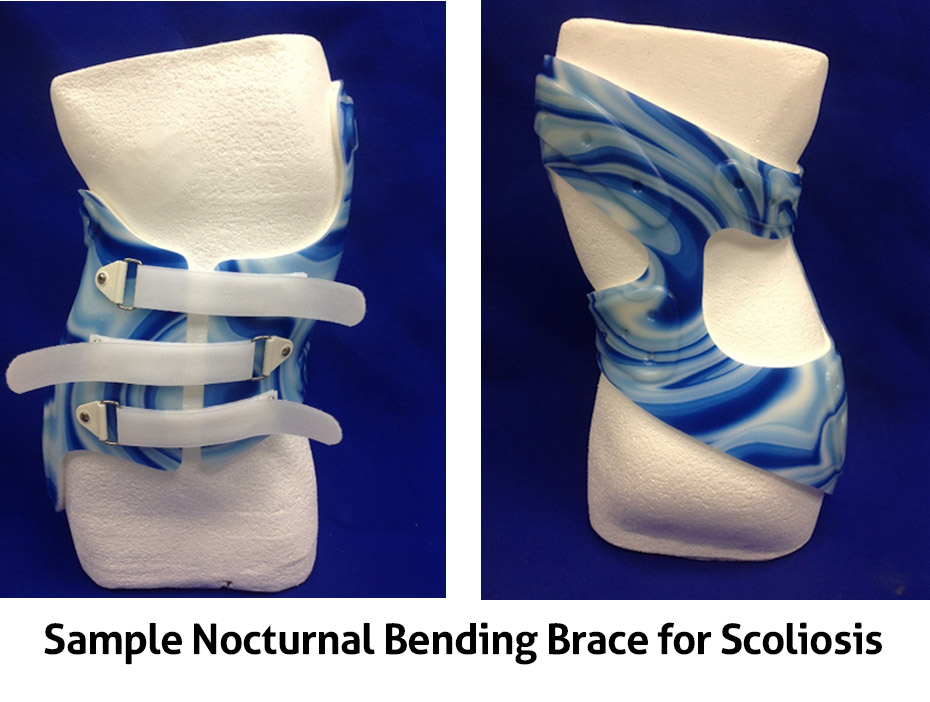
Through years of development Owens Carolina, has spent time learning from the past. Since the 1940’s a great deal of research has been dedicated to scoliosis bracing. It has always been known that scoliosis is a 3 dimensional issue, not a 2 dimensional problem.
Spondylosis Braces and Bracing
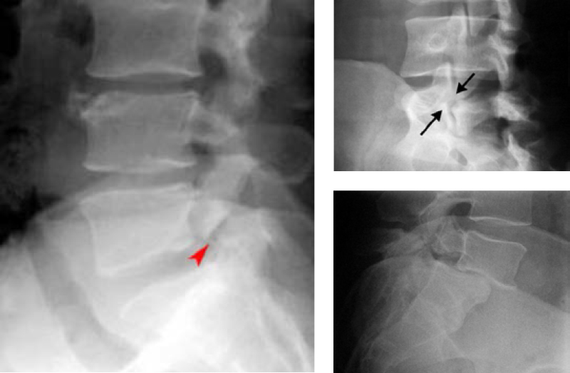
Spondylosis is a fairly common injury to the spine. It can go by many names. Pars fracture, pars defect, scotty dog fracture (I’ll explain), just to name a few. Basically, you have fractured the pars interarticularis bone in your spine. When this fracture is viewed on the x-ray, you can see the silhouette of a scotty dog, (top right) and it appears that the neck is broken. For each vertebra there are 2 pars bones. It is possible you fractured one of them, and it is possible you fractured both. The most common vertebra that has this injury is your 5th lumbar. This is the last mobile vertebra in your spine and sits on top of the sacrum (your pelvis). If your x-ray looks like the one on the left, this is the reason you are in our office. As you can see, the pars interarticularis is fractured. The good news is the vertebra has not shifted forward. If we can hold everything in place for a few months, you will probably heal, and no further intervention will be necessary. If your x-ray looks like the one on the bottom right, you are probably not getting a brace because you are scheduled for surgery. It is possible your surgeon may order a brace after the surgery. This is a good thing because it will make you feel better and is only needed for a few months while you heal.
The typical person that gets this brace is young, 13 to 18 years old. They are an athlete. Couch potatoes do not have this problem. It is possible they have had back pain for several months, but because body pain is associated with hard training, this sometimes gets the credit for the discomfort. They also know that if they tell the coach, they may get benched. Eventually, the parents seek help and the orthopedist immediately suspects this problem. Many years ago, a soon to retire orthopedic surgeon told me, “if I can get both sides of that bone in the same room together, they will heal.” If the proper brace is worn and activities are halted, the young athlete will be back on the field in the future.
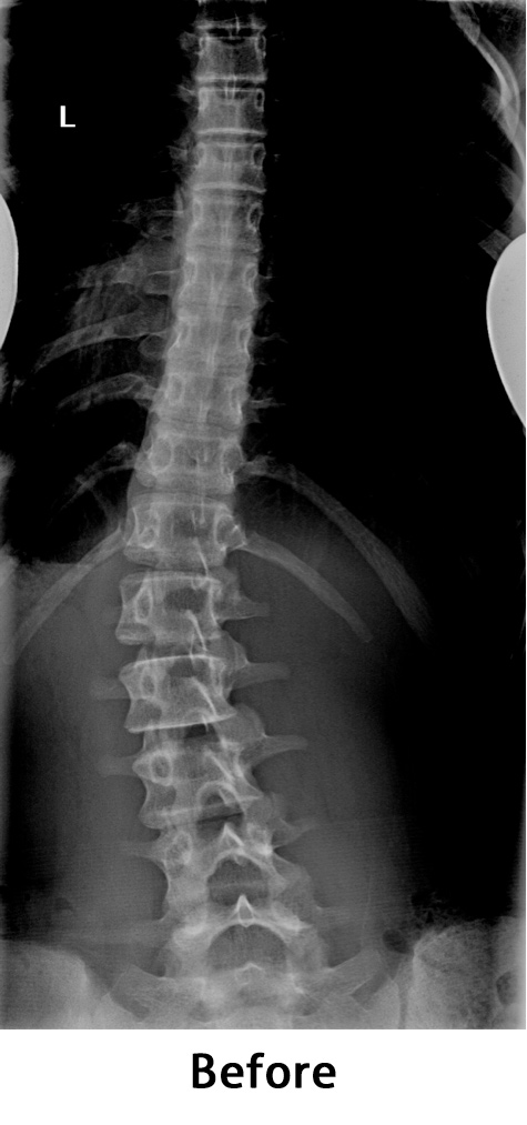
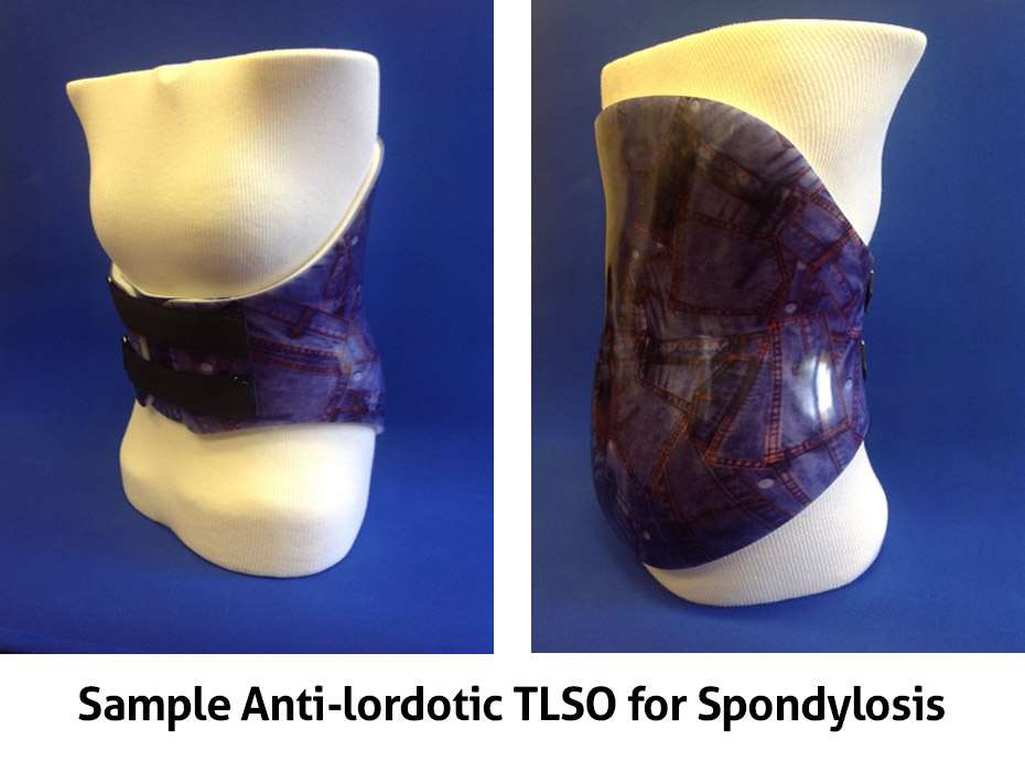
Important, when being braced the spine must be forced back. There must be pressure on the stomach and nothing touching the back. This is often described as lumbar flexion or anti-lordotic. The terms mean the same. When properly fit there should be space between the back of the brace and the lower back. As you can see by the x-ray we want to create a force that pushes the lumbar spine from the front to the back. If we are to push the spine to the back, there must be a void area there. The LSO or TLSO with 0 degrees of lordosis is the required brace and is usually the first choice.
Compression Fractures
Compression fractures are a common form of injury to the spine. There can be many causes. Auto accidents, falls and certain diseases to name a few. As you can see by these examples, the vertebrae have been compressed. This happens at the moment of impact and the vertebra are crushed. These types of injuries are very painful, but generally do not result in spinal cord injury. The vertebral body is the main load bearer of the spine. Therefore, it can be more painful when sitting or standing than laying.
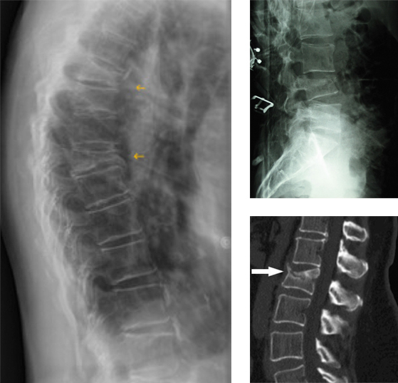
When treating the fracture with a brace, there are just a few goals. The first is to unload the fracture site. This is achieved by forcing the spine into extension. By making you stand up straighter, weight is transferred to the articular facets on the back, and the brace on the front. Moving the spine to this position is painful, but will make you feel much better. The next goal is to aid the healing process. By extending the spine, the vertebrae will heal less deformed. The spine is going to heal, braced or not. By wearing the brace, immediate pain is reduced, deformity is reduced and future potential issues can be avoided. There are many types of braces used in the treatment of compression fractures. We will work with you and your physician to provide the best option and get you through this event in your life.